
William N. Ross, Ph.D., of New York Medical College and the Marine Biological Laboratory, uses imaging techniques to study synaptic integration and plasticity in dendrites — branched extensions of neurons that receive in coming signals from different parts of the brain. To learn more about the calcium and sodium interactions that take place in neurons, Ross and his colleagues developed a technique for imaging both sodium and calcium ion signals in quick succession. Incorporating high intensity LEDs from Prizmatix into their imaging setup made it possible to achieve the fast excitation light switching and high signal-to-noise ratios needed to capture small, fleeting changes in fluorescence intensity.
LED advantages
To image calcium and sodium simultaneously, the researchers loaded calcium and sodium fluorescence indicators into a single neuron and then quickly switched between the excitation wavelengths for each indicator at 500 Hz. The use of LEDs to provide the excitation light was advantageous because they could be turned on and off instantaneously without a shutter, which cannot respond this fast and can introduce vibrations. To take full advantage of the fast LED switching, the researchers synchronized it to the frame rate of their high-speed camera. “This rapid switching let us alternately illuminate different indicator dyes very rapidly,” said Ross.
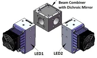
The outputs of the two Prizmatix LEDs are combined with a dichroic mirror. Because the LEDs are modular, the researchers easily, and cost-effectively, incorporated various LED combinations into the excitation port of their fluorescence microscope.
“We found it useful that the Prizmatix LEDs could be controlled by gating, a variable intensity dial, or analogue input. There was no extraneous computer control, which makes some other systems more expensive without providing a real advantage.”
Prizmatix offers LEDs that have among the highest intensity of any on the market — five to ten times brighter than arc lamps in the narrow wavelength bands used for exciting fluorescent dyes. This high intensity allowed the researchers to acquire better measurements since, in the small compartments of single neurons, the signal-to-noise ratio is proportional to the square root of the light intensity.
Through test experiments, the researchers showed that their method produced an improved signal-to-noise ratio compared to approaches other researchers had used for simultaneous imaging of sodium and calcium ions. While conducting these experiments, the researchers realized that the LEDs came with another advantage: the cooling fans turned on only after a few second delay. Since this delay was usually longer than the duration of the measurements, there was no disturbance from fan vibrations.
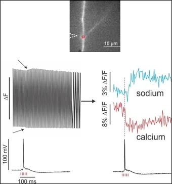
The top image shows part of a pyramidal neuron dendrite in a brain slice from the rat hippocampus with a region of interest (red) marked near a branch point. The patch electrode on the soma contained 200 µM ANG-2 (sodium indicator) and 200 µM bis-fura-2 (calcium indicator). The recording on the bottom left shows the envelope of the combined signal from the region of interest, with the two arrows indicating changes resulting from an extracellular stimulus. The recording on the right shows the separated signals, which go in opposite directions as the fluorescence changes from the two indicators either increase (ANG-2) or decrease (bis-fura-2) with increasing ion concentrations. The black trace on the bottom shows the electrical recording from the soma taken at the same time as the optical traces. Figure courtesy of William N. Ross.
Precision measurements
“The Prizmatix LEDs met all our expectations and have been very reliable,” said Ross. “In fact, the intensity of the LEDs was much higher than we originally expected, allowing us to make more precise measurements.”
After experiencing success with using one pair of LEDs, the researchers designed new experiments that take advantage of Prizmatix LEDs that optimally excite other indicator dyes. They now have four different LEDs, covering different spectral ranges, that can be used for different experiments.
“I found the people at Prizmatix very helpful. They clearly understood what I was trying to accomplish with the LEDs,” said Ross. “They let me try different components before purchasing them and supplied all the pieces needed to connect the LEDs to our microscope.”
Research paper: Miyazaki K. Ross WN. Simultaneous Sodium and Calcium Imaging from Dendrites and Axons. eNeuro 14 October 2015, 2 (5) ENEURO.0092-15.2015
 UHP-LED for Sodium and Calcium Imaging
Prizmatix Products for Research
In this research Dr. Ross used Prizmatix Ultra High Power Collimated LEDs (UHP-T-LEDs) at wavelengths 385 nm, 460 nm and 520 nm. Since then Prizmatix improved and expanded UHP-T-LED product lines and now we offer more advanced models. Currently main product lines are:
UHP-T-MP – Most powerful collimated LED with rectangular shape LED emitter (about 3x4mm). Providing rectangular beam for applications such as microplate illumination.
UHP-T-DI - Powerful collimated LED with square shape LED emitter (3x3mm). Providing square beam for application such as epifluorescence microscopy, petri dish illumination, optional 5mm core light guide coupling.
UHP-T-EP – Powerful and high brightness collimated LED with square shape LED emitter (2x2mm). Providing square beam for application such as epifluorescence microscopy, DMD, Optogenetics, optional 3mm core light guide coupling.
UHP-T-SR – Ultra Bright collimated LED with small LED emitter (several sizes available). Providing extra brightness for application such as epifluorescence microscopy, Optogenetics, optional 3mm core light guide coupling and fiber coupling.
Advantages of UHP-T series:
Location of high current LED driverIn UHP-T-LED the high current LED driver is located inside LED head rather than in controller. In similar products from other vendors the high current driver located in the controller box. When high current flows from controller to LED head high RFI/EMI interference may disturb delicate measurements. In UHP-T-LED models the LED head is grounded and serve as Faraday cage reducing the RFI/EMI interference.
Optically Isolated TTL and Analog InputsThe controller of UHP-T-LED features, as standard, TTL input for very fast triggering (or ON/OFF strobing) of LED light without need of a shutter. The Analog input provides simple way to control the LED light power from computer. Both inputs are independent and optically isolated to eliminate ground loops.
Low Optical Noise OptionMost modules can be purchased with Low Noise (-LN) option. The low optical noise light source is very important in experiments involving such measurements as cell Sodium and Calcium imaging. In many preparations the change of fluorescence signal may be just few percent. If the excitation light source exhibits high intensity fluctuations the small fluorescence changes may be obscured. UHP-T-LED-LN has intensity fluctuations of less than 0.01% RMS between DC to 1MHz, enabling detection of small changes in fluorescence intensity.
The contemporary LED light source for Sodium and Calcium imaging is UHP-T-EP-LN. This model provides besides powerful illumination low RFI/EMI interference, low optical noise and convenient TTL and Analog inputs. All these features are important in LED light-source for Ephys rig environment.